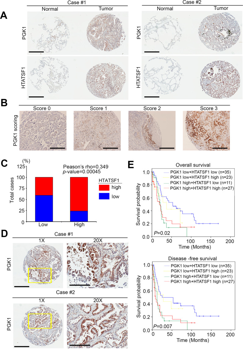
Fig. 7. PGK1-HTATSF1 expression as a prognostic factor for clinical lung cancer patients.
A PGK1 and HTATSF1 protein expression in the paired normal and tumor tissues derived from clinical lung cancer patients. Statistical significance was analyzed by a paired t-test. Scale bar: 400 μm. B IHC staining for the PGK1 protein. The intensity of IHC staining was scored as a range from 0 to 3. Scale bar: 50 μm. C Quantification of HTATSF1 expression by immunohistochemistry analysis of lung cancer specimens by each corresponding clinical parameter. D PGK1 protein expression in several tumor tissues derived from clinical lung cancer patients. Scale bar: 400 μm. 20x: 200μm.E Kaplan–Meier analysis of the overall survival and disease-free survival probabilities of clinical lung cancer patients according to the intensity (low = 0 and 1, high = 2 and 3) of IHC staining for the PGK1 protein combined with the HTATSF1 protein level.

