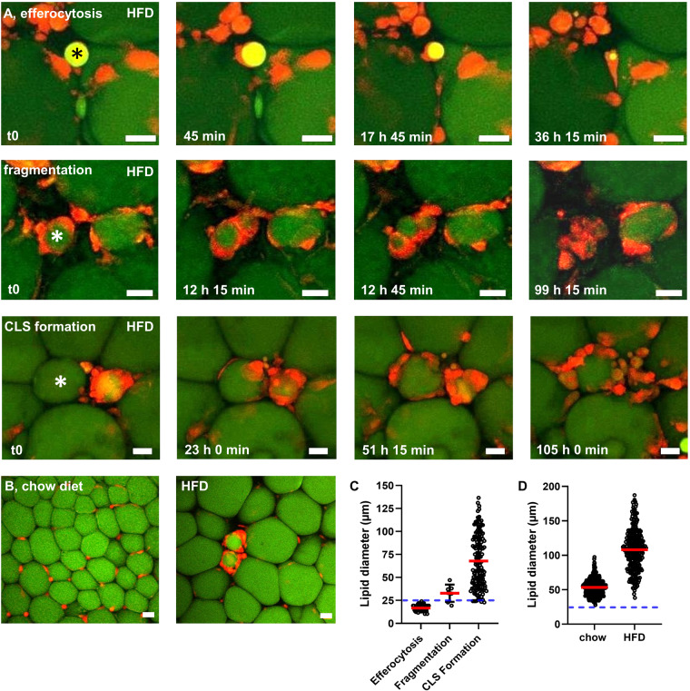
Fig. 1. Live-imaging indicates a size threshold for efferocytosis of lipid remnants.
Live-imaging of AT explants of HFD-fed MacGreen mice (green: BODIPY-stained lipids, red: ATMs, movies provided as online supplement). A Degradation of adipocyte remnants in AT explants occurs in three distinct ways: efferocytosis (upper row), fragmentation (middle row), or CLS formation (lower row). B Representative overview images of living AT of chow-fed and HFD-fed mice. C Quantification of lipid diameter associated with either efferocytosis, fragmentation, or CLS formation (188 registered events in 40 movies from 6 independent experiments). D Quantification of lipid diameter in adipocytes of chow-fed or HFD-fed littermates (N = 3). Asterisks mark lipid remnants degraded by ATMs. Blue line indicates the threshold for efferocytosis. Scale bars = 25 µm.

