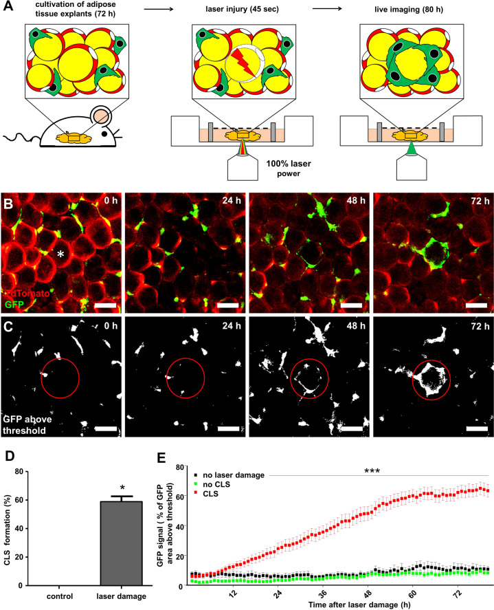
Fig. 2. Model of targeted adipocyte death by laser injury in living AT.
A Scheme of laser injury protocol and B formation process of CLS after adipocyte death in AT explants from lean double reporter mice (Csf1r-eGFP x AdipoqCreERT2:TDTO mice). Movie provided as supplemental online material. C, E GFP fluorescence quantification in proximity to the targeted adipocyte (E, no laser damage n = 13; no CLS n = 23; CLS formed n = 35; N = 5). D CLS formation after laser injury (N = 5). Scale bars = 50 µm. *p < 0.05 and ***p < 0.001. Data presented as mean ± SEM.

