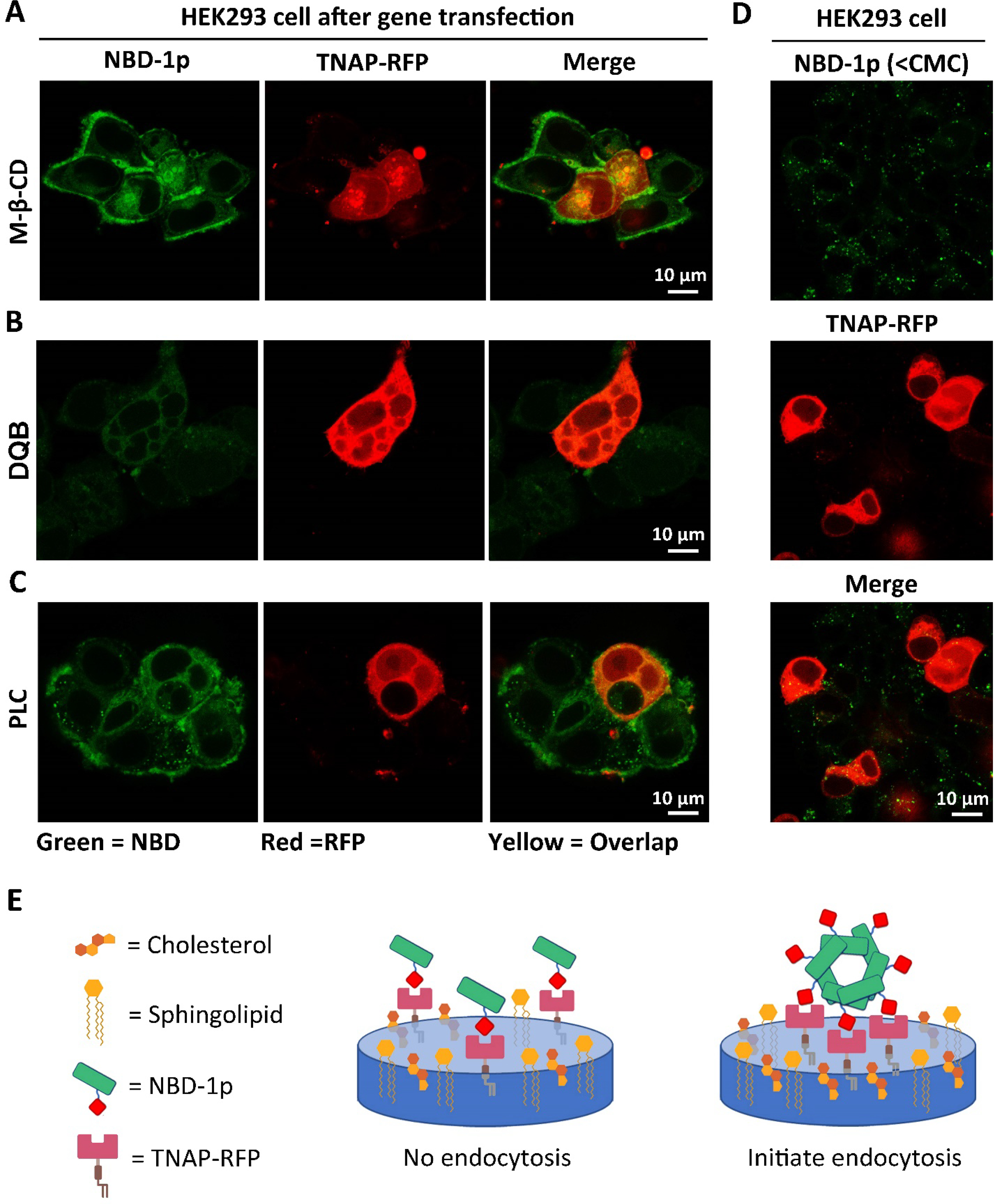
Figure 2.

NBD-1p nanoparticles associate with the TNAP in lipid rafts followed by CME. (A-C) Confocal fluorescence images of HEK293_TNAP-RFP cells incubated with NBD-1p (200 μM, 12 h) after the pretreatment of (A) caveolin-dependent endocytosis inhibitors (M-β-CD), (B) TNAP inhibitor (DQB), and (C) phospholipase C which removes the GPI-anchored TNAP on plasma membrane. (D) Confocal fluorescence images of HEK293_TNAP-RFP cells incubated with NBD-1p (50 μM, 12 h) below CMC. (E) Illustration of that the assemblies of phosphopeptide rather than individual molecules initiate CME.
