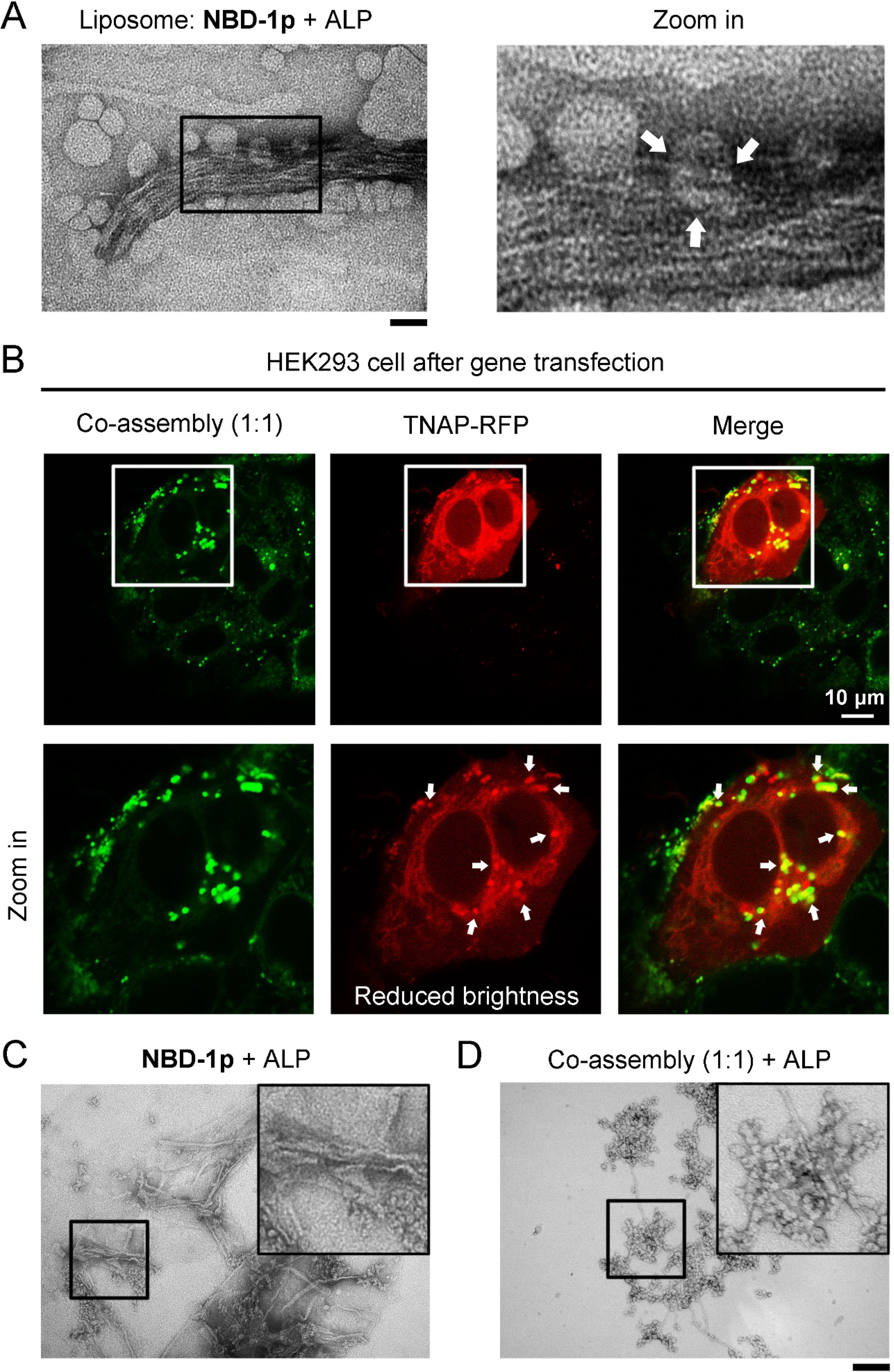
Figure 4.

The co-assembly of NBD-1p and NBD-(D)Sp shows less efficient endosomal escape. (A) TEM image of liposomes carrying NBD-1p and ALP (1 U/mL, 37 °C, 6 h). Broken liposomes are indicated by arrows. Scale bar = 100 nm. (B) Confocal fluorescence images of HEK293_TNAP-RFP cells incubated with the mixture of NBD-1p (100 μM) and NBD-(D)Sp (100 μM) for 12 h. The peptides trapped in endosome are indicated by arrows. (C) TEM images of NBD-1p (200 μM) and (D) the co-assemblies of NBD-1p (100 μM) and NBD-(D)Sp (100 μM) after the addition of ALP (0.1 U/mL, 37 °C, 3 h) for partial dephosphorylation. ALP yields more nanofibers in (C) than in (D). Scale bar = 100 nm.
