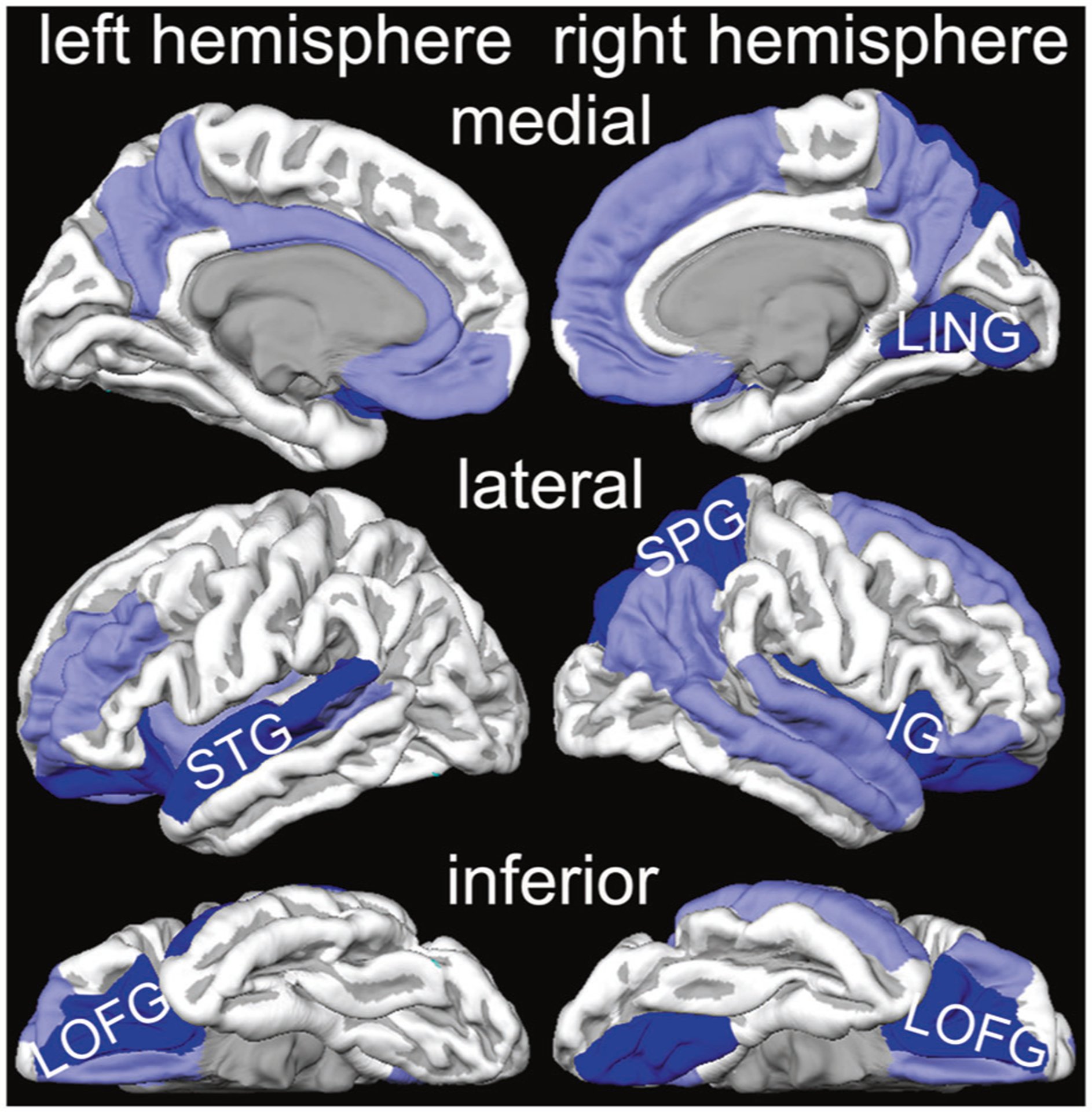
Fig. 1. Cortical volume differences between PTSD and control subjects.

Light blue indicates regions with smaller volume in PTSD group. Dark blue indicates regions which are smaller in PTSD group, and their volumes are negatively associated with harmonized PTSS severity scores.
