FIGURE 1.
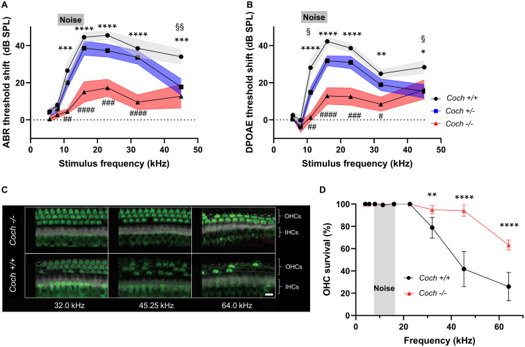
Absence of cochlin reduces the level of cochlear dysfunction and sensory cell damage after acoustic injury. Six-week-old mice of each genotype were exposed to 8–16 kHz noise for 2 h at 103 dB SPL. (A) ABR and (B) DPOAE threshold shifts 2 weeks after noise trauma demonstrate profound mid-to-high frequency hearing loss in wild-type Coch+/+ mice, and statistically significant lower threshold shifts in Coch–/– mice. A trend toward lower threshold shifts is observed in heterozygous mice, which reaches statistical significance at 11.33 kHz (DPOAE) and 45.25 kHz (ABR and DPOAE). The gray rectangle indicates frequency of noise band. Data are shown as group means ± standard error of the mean; N = 10 Coch+/+, N = 11 Coch+/–, and N = 11 Coch–/– animals. *P < 0.05, **P < 0.01, ***P < 0.001, or ****P < 0.0001; asterisks: Coch+/+ vs. Coch–/–, #: Coch+/–. Coch–/–, §: Coch+/– vs. Coch+/+. (C) Representative cochlear whole mounts from Coch+/+ and Coch–/– mice 2 weeks after acoustic trauma. IHC, inner hair cells. OHC, outer hair cells. Green = myosin 7A, white = phalloidin. Scale bar: 10 μm. (D) Cochleogram showing fewer missing outer hair cells in Coch–/– mice 2 weeks after acoustic trauma. Data are shown as group means ± standard error of the mean; N = 4 Coch+/+ and N = 4 Coch–/– mice. **P < 0.01 or ****P < 0.0001.

