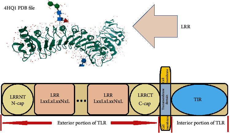
Figure 3.

The structure of TLR and the related domains. The Ls in LxxLxLxxNxL depict hydrophobic core built of β-strands, and the N depicts the Asparagine network. The two-solenoid LRR structure shows the 3D structure of loops, helices (convex surface), and β-strands (concave surface) (4HQ1 PDB file) [87].
