
Figure 2.
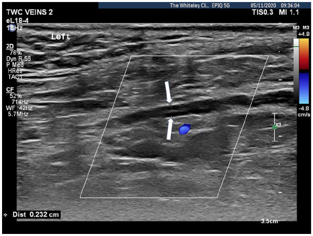
Duplex ultrasound showing thrombosed and left testicular vein with no flow (cursors showing reduced diameter of thrombosed vein shown by white arrows).
Official websites use .gov
A
.gov website belongs to an official
government organization in the United States.
Secure .gov websites use HTTPS
A lock (
) or https:// means you've safely
connected to the .gov website. Share sensitive
information only on official, secure websites.
Duplex ultrasound showing thrombosed and left testicular vein with no flow (cursors showing reduced diameter of thrombosed vein shown by white arrows).