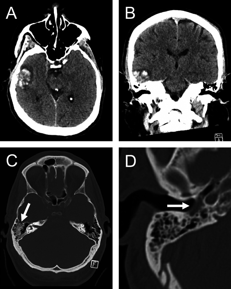
Figure 1.
Axial (A) and coronal (B) unenhanced CT of the head showing an acute right lateral temporal hemorrhagic contusion. Though no skull fracture was visualized, partial opacification of the right mastoid air cells (C, arrow) and right tympanic cavity (D, arrow) by fluid suggests possible injury to adjacent bone.

