Fig. 4.
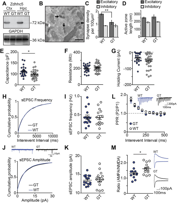
Zdhhc5 regulates excitatory synapse density in vivo. (A) Western blots of cortical (Ctx) and hippocampal (Hpc) lysates from Zdhhc5-GT (gene trapped) mice and age-matched littermate controls showing the absence of Zdhhc5 expression in Zdhhc5-GT mice. (B) Representative immunogold-electron microscopy images showing excitatory symmetric synapses (black arrows) and inhibitory asymmetric synapses (white arrows). Scale bar: 500 nm. (C) Zdhhc5-GT mice exhibit a significant reduction in the density of excitatory synapses, with no change in inhibitory synapses. n=3 mice per genotype. **P<0.01 [two-way ANOVA, significant interaction between genotype and synapse type, F(1,8)=8.753, P=0.0182, Bonferonni's test post hoc, n=3 mice per genotype]. (D) There is no significant difference in the active zone between Zdhhc5-GT and WT littermates. (E) CA1 pyramidal neurons from acute Zdhhc5-GT brain sections exhibit lower capacitance than pyramidal neurons from WT mice (n=24 WT neurons; 4 mice and 31 Zdhhc5-GT neurons; 5 mice) *P=0.0127 [unpaired two-tailed t-test, t(53)=2.579]. (F,G) There is no significant difference between WT and Zdhhc5-GT mice in either resistance (F) or holding current (G). (H–J) There is no significant difference of sEPSC frequency (H,I), sEPSC amplitude (J,K), or paired-pulse ratio (L). (M) CA1 pyramidal neurons from Zdhhc5-GT brain slices exhibit a significantly higher AMPAR to NMDAR current ratio than WT slices. n=12 WT neurons; 3 mice and 13 Zdhhc5-GT neurons; 3 mice. *P=0.0363 [unpaired two-tailed t-test, t(23)=2.224]. Insets in L, J and M show representative current traces from each experiment. All data is from comparisons of Zdhhc5-GT with age-matched littermate controls and is shown as mean±s.e.m.

