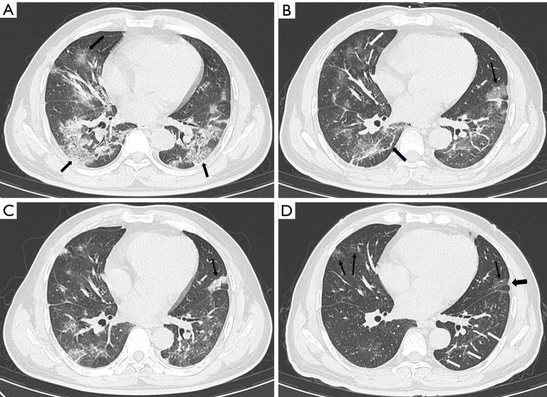
Figure 3.
Serial chest CT scans of a 48-year-old female were graded as severe. (A) The scan obtained at the onset of symptoms showed GGO (thin arrow) with a “crazy-paving” pattern and air bronchogram (black arrow). (B) The scan obtained 7 days after the onset of symptoms showed evolution to consolidation (white arrow), and a mixture of GGO and consolidation (thin arrow) with increased density and decreased extension. (C) The scan obtained at discharge showed a mild decrease in extension than previous scans and minimal absorption; an air bronchogram was also observed (black arrow). (D) The scan at 8 weeks after discharge showed the evident absorption of previous abnormalities and multiple GGOs (white arrows) of decreased density and increased extension. CT, computerized tomography; GGO, ground-glass opacity.

