Figure 3.
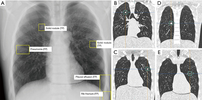
Images of a chest phantom with four solid nodules in both lungs. (A) PA radiograph with the marked findings of the AI based CAD system. Two solid nodules were correctly identified by the software (TP). Rib fracture, pneumonia and pleural effusion were false FP. (B,C,D,E) Coronal CT images of the same chest phantom showing the four nodules in the left lung. The smaller nodule on the left and the more centrally located nodule on the right were missed by the software. FP, false positive; TP, true positive; PA, posteroanterior; CAD, computer-aided-diagnostic.

