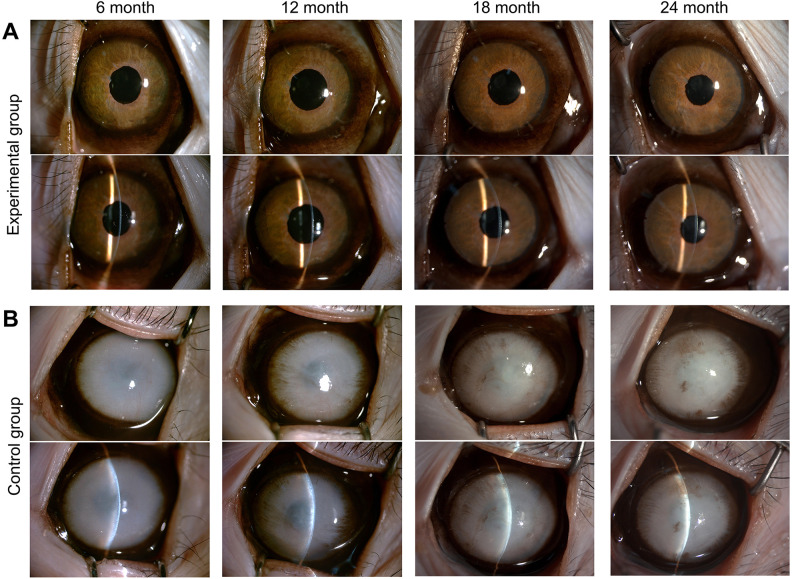
Figure 2.
Clinical observation of the experimental group and control group during 2 years. (A) In the experimental group, the corneas remained transparent, and almost no keratic precipitates or corneal neovascularization appeared. (B) In the control group, the corneas remained opaque with edema and as a consequence the iris could not be seen. Monkey 1 and 2 are shown here. The other three monkeys are shown in Supplemental figure 2.

