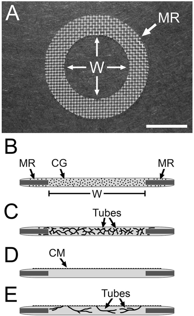
Figure 2.

Nylon mesh ring system used to study HUVEC tubulogenesis. (A) Image of a nylon mesh ring (MR) on which the collagen gel is cast. HUVEC tubes that form in the collagen gel are viewed in the central “window” region (W) of the gel. (B–D) Side-view diagrams of the setup for multiaxial (B, C) and monolayer-sprout (D, E) culture modes. (B) HUVECs (black dots) are dispersed in the collagen gel (CG). Nylon mesh ring (MR) and window region (W) of the collagen gel are indicated. (C) The dispersed cells organize into multiaxial HUVEC tubes in response to T-medium. In monolayer-sprout cultures (D), a confluent monolayer (CM) of HUVECs on top of the collagen gel is induced to sprout into the gel (E). Scale bar size: A = 5 mm. Abbreviations: HUVEC, human umbilical vein endothelial cell; T-medium, tubulogenesis medium.
