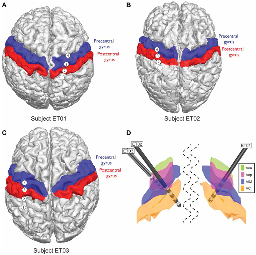
Fig. 1. Patient-specific magnetic resonance imaging–computed tomography reconstruction and segmentation of cortical and thalamic regions in ET closed-loop patients.

(A) Subject ET01, (B) subject ET02, and (C) subject ET03 cortical segmentations. Red, postcentral gyrus; blue, precentral gyrus, known as primary motor cortex (M1). (D) Subcortical thalamic segmentation with overlaid DBS lead positioning based on surgical planning and all normalized to an MNI brain. The structures shown are ventralis oralis anterior (Voa; green), ventralis oralis posterior (Vop; magenta), ventralis intermediate nucleus (VIM; blue), and nucleus ventralis caudalis (VC; orange).
