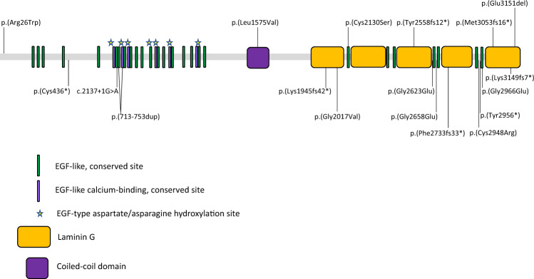
Fig. 1. Schematic diagram showing the protein domain structure of EYS.
The protein consists of EGF-like and EGF-like calcium binding domains at the N terminal end, a coiled coil domain and 5 laminin G binding domains at the C-terminal end. The mutations identified in our cohort are marked, those novel to this study are shown above the structure and those reported previously are shown below (see Table 3 for details).

