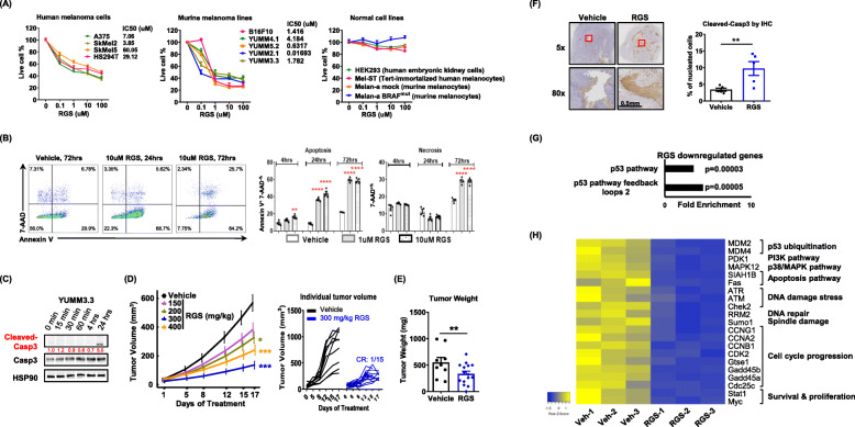
Fig. 1.
Rigosertib induces melanoma cell death and inhibits melanoma tumor growth in vivo. a IC50 determined by CellTilter Blue assay at 72 h post RGS treatment. The melanoma and normal cells were seeded in 96-well plate with 3000 cells per well. b Dot plots and quantified results of the Annexin V+ and 7-AAD+ cell percentage in YUMM3.3 culture with or without RGS treatment. c Whole-cell extracts were harvested and immunoblotted. HSP90 was used as a loading control for densitometry quantification (red numbers). d, e Tumor volume and weight of YUMM3.3 melanomas in C57BL/6 female mice. Daily RGS treatment starts at day 10 post tumor cell inoculation. CR, complete regression. f Representative images and quantitative results of cleaved-caspase3 protein levels observed from day 17 post RGS treatment (300 mg/kg) of YUMM3.3 tumors by immunohistochemistry. g Gene ontology enrichment analysis of gene targets downregulated in YUMM3.3 tumors post 17 days of RGS treatment (300 mg/kg). Results for Fold Enrichment > 3 and FDR p < 0.05 are displayed. h Heatmap summarizes 21 p53-associated targets from a total of 1495 genes screened that were significantly downregulated due to RGS treatment. a-f pooled data obtained from at least two different experiments (n = 4 ~ 15) are shown. g, h were triplicates

