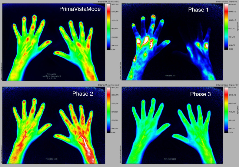
Figure 1.
FOI images in PrimaVistaMode and phases 1–3 in a patient with active, early seropositive rheumatoid arthritis. Early and strong enhancements within MCP joints in phase 1. Furthermore, moderate enhancement of grade 2 within right PIP joints in phase 2. FOI, fluorescence optical imaging; MCP, metacarpophalangeal; PIP, proximal interphalangeal.

