Figure 3.
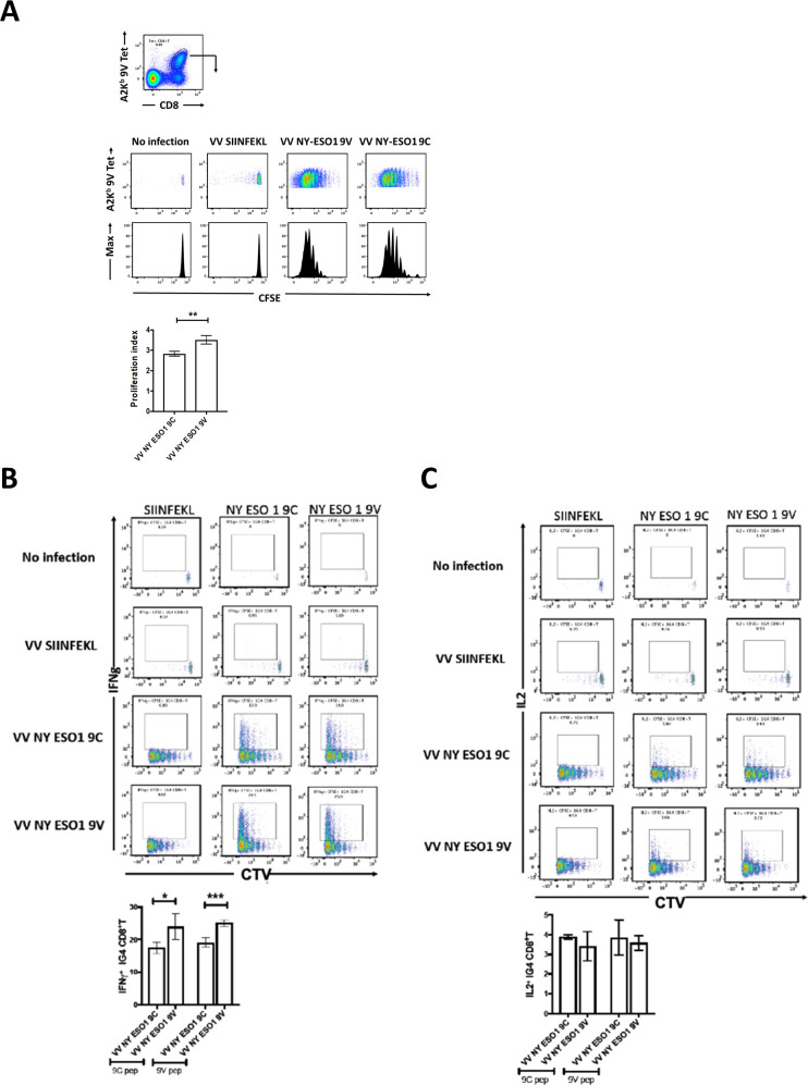
m1G4 CD8 +T cells show antigen specificity. (A) HHD mice were adoptively transferred via intravenous injection with 2×10⁶ CellTrace Violet (CTV)-labeled naive CD8 +T cells from 1G4 mice and left as uninfected or infected intravenously with 1×10⁶ pfu of rVV expressing full length NY-ESO-1 containing either wild type SLLMWITQC or altered peptide ligand SLLMWITQV. As control, rVV expressing SIINFEKL (from chicken ovalbumin) peptide was used. CTV dilution profiles of splenic m1G4 cells were analyzed at day three postinfection having been stained with A2Kb/NY-ESO-1157–165V tetramer. (B) Splenocytes from the infected mice were restimulated ex vivo with SIINFEKL, SLLMWITQC or SLLMWITQV peptides and IFN-γ expression or (C) IL-2 expression were analyzed by intracellular cytokine staining (ICS) and CTV dilution on day 3. For each panel, representative FACS plots are shown in the top panel and cumulative data for mouse groups (n=5) are plotted below. *P<0.05, **p<0.01, ***p<0.001 in Student’s t-test with GraphPad software. FACS, fluorescence-activated cell sorter; IFN-γ, interferon-γ; IL-2, interleukin 2.

