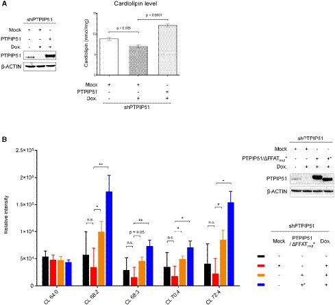
Figure 5. Mitochondrial CL levels mediated by PTPIP51.

- CL levels after PTPIP51 depletion and reconstitution were monitored. A doxycycline‐induced shRNA expression system was used to deplete the PTPIP51 protein in HeLa cells. Left, Western blot analysis of HeLa cells in which PTPIP51 was depleted and restored. β‐ACTIN was used as the loading control. Right, the mitochondrial CL level (nmol/mg) according to PTPIP51 expression. To reconstitute PTPIP51, the full‐length PTPIP51 gene was transiently expressed. The data are presented as the mean ± SD of technical triplicate experiments from one representative experiment (n = 3), and P‐values calculated using Student’s t‐test are shown.
- Quantification of CL in mitochondria using lipidomic analysis after PTPIP51 depletion and reconstitution. Color codes for each sample were presented in the lower right subpanel. Experiments were technically repeated three times, and the results are represented as the means ± SD of technical replicates (n = 3). *P < 0.05; **P < 0.01 (Student’s t‐test). Quality control (QC) samples, which were pooled identical aliquots of the mitochondria samples, were measured four times throughout the run for data reproducibility. Upper right, Western blot analysis of HeLa cells in which PTPIP51 WT or ΔFFAT mutant was depleted and restored. β‐actin was used as a loading control.
