Figure 4.
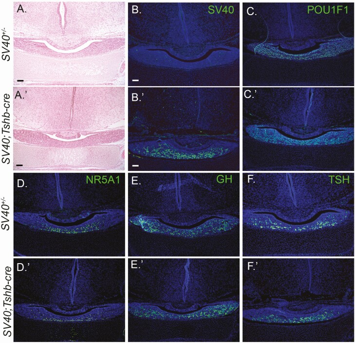
SV40; Tshb-cre mice have normal pituitary morphology and cell specification at birth. Coronal sections of P1 pituitaries from SV40 positive, cre negative (A-F) and SV40; Tshb-cre mice (A′-F′) were stained with H&E and various antibodies (N = 3 individuals/genotype). The magnification is 100× with 100 μm scale bar. H&E staining (A, A′) and immunostaining for SV40 (B, B′), POU1F1 (C, C′), NR5A1 (D, D′), GH, (E, E′), and TSH (F, F′). DAPI staining (blue) of cell nuclei B-F′.

