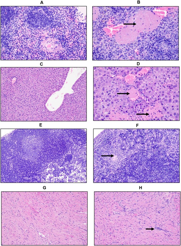
Figure 4.
Histopathologic changes in IFNAR-/- mice infected with AHFV. Groups of six mice were infected IP with 1000 LD50 (400 TCID50) and euthanized at 2 dpi and 4 dpi for organ harvest. (A and B) H&E stain of spleens from mice at 4 dpi (A, Mock 400×; B, AHFV infection 400×). We observed a reduction in the white pulp and expansion of the red pulp in infected animals indicating lymphoid depletion. There was neutrophil infiltration and vascular occlusion by a fibrin thrombus (arrow). (C and D) H&E stain of livers from mice at 4 dpi (C, Mock 200×; D, AHFV infection 400×). Hepatocytes were swollen and contained lipid micro-vesicles. There was an increase in sinusoidal inflammatory cells and, along the bottom of the frame, a focus of necrosis (bottom arrow). Fibrin thrombi occluded adjacent vessels (top arrow). (E and F) H&E stain of cervical lymph nodes from mice at 4 dpi (E, Mock 200×; F, AHFV infection 200×). Lymph nodes from infected animals exhibited tremendous lymphocyte apoptosis, infiltrating neutrophils and occasional fibrin thrombi (arrow). (G and H) H&E stain of heart from mice at 4 dpi (E, Mock 200×; F, AHFV infection 200×). Increased cellularity due to interstitial inflammation was seen in infected animals (arrows).

