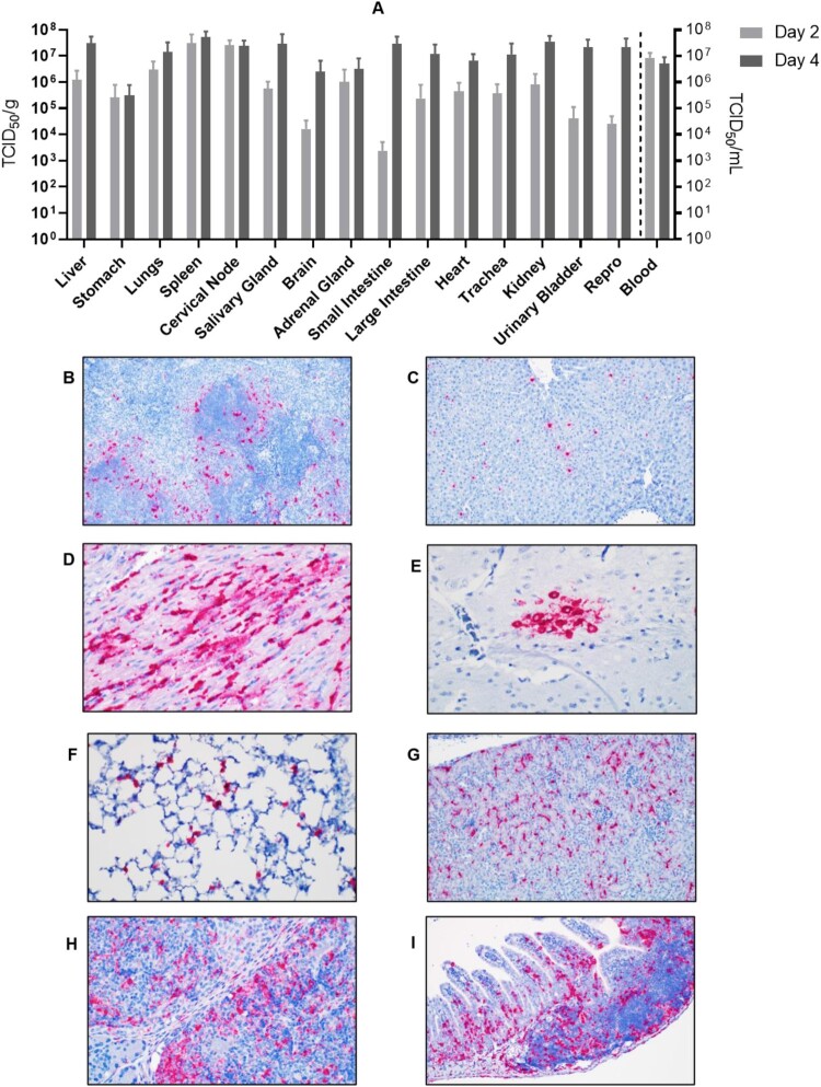
Figure 5.
Tissue tropism of AHFV in infected mice. Groups of six mice were infected with 1000 LD50 (400 TCID50)/mouse via the IP route. The groups were euthanized on 2 and 4 dpi and organs were harvested to determine virus load. (A) The viral load in tissue is presented in TCID50/g; viremia is presented as TCID50/ml. (B–I) In-situ hybridization of AHFV genome in organs of infected mice. (B) Spleen 2 dpi; AHFV RNA was present at the marginal zone where the red and white pulp interact (200×); (C) Liver 4 dpi; AHFV RNA was located within individual mononuclear cells (Kupffer cells; 200×); (D) Heart 4 dpi; AHFV RNA corresponded with areas of inflammation (400×); (E) Brain 4 dpi; AHFV RNA was focally present within a cluster of neurons in the grey matter of the temporal lobe (400×); (F) Lungs 4 dpi; AHFV RNA was multifocally present throughout alveolar septa, presumably within macrophages (400×); (G) Kidney 4 dpi; AHFV RNA was detected within the interstitial tissue of the kidney (200×); (H) Cervical lymph node 4 dpi; AHFV RNA was located within mononuclear cells of the subcapsular sinuses and throughout lymphoid follicles (400×); (I) Small intestine, gut associated lymphoid tissue 4 dpi; AHFV RNA was present in GALT and lamina propria of intestinal villi (200×).

