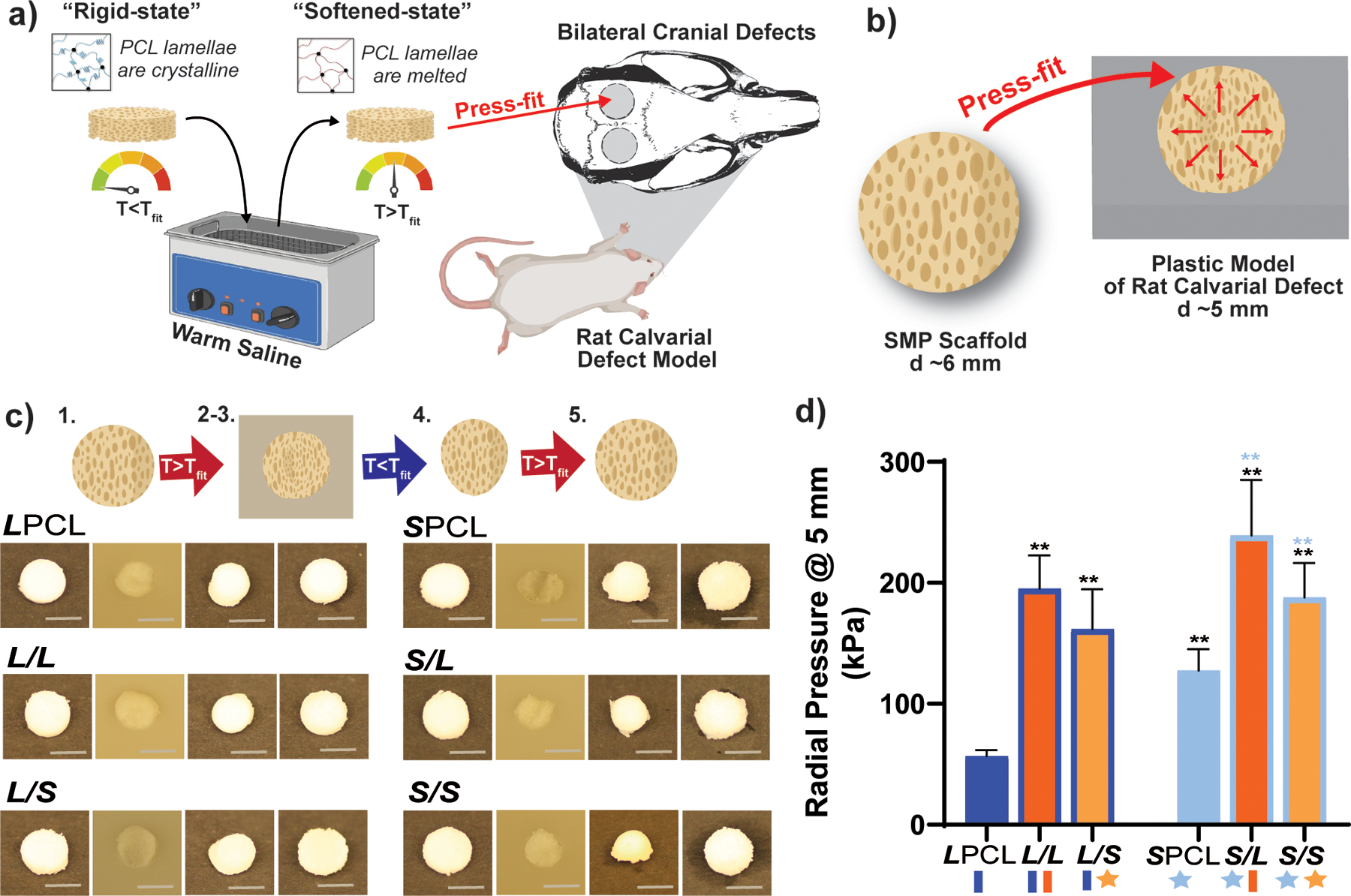
Figure 5.

(a) shape memory testing was performed to mimic a bilateral rat calvarial defect model in vivo study. (b) scaffolds were designed to be slightly larger than the cranial defect, so the warm scaffold will exert a force on the defect edges, as shown in the schematic. (c) all compositions were able to be press-fitted into a plastic model defect and demonstrated excellent shape fixity/recovery. protocol: following submersion in saline at tfit for 1 minute [step 1], all scaffolds were successfully press-fitted into defects (i.e. expanded via shape recovery to fill the defect) [step 2]. after just 2 minutes within the defect, scaffolds returned to their relatively rigid state (i.e. underwent shape fixation in new shape within the defect) [step 3]. next, scaffolds were removed from the defect and allowed to sit for 2 min (to determine shape fixity) [step 4] and reheated at tfit in saline for 1 minute (to determine shape recovery) [step 5]. (d) radial expansion pressure tested at tfit; *p < 0.05, **p < 0.01.
note: black color-coded statistics are compared to lpcl and “blue color-coded” statistics are compared to spcl.
