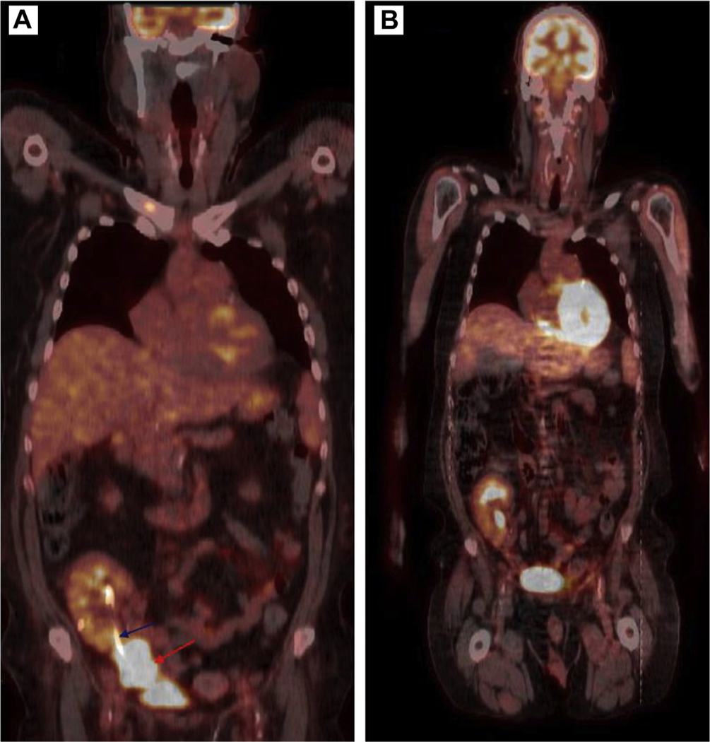
Figure 2.
Positron Emission Tomography Scan Image at Diagnosis, Prior to Initiation of Treatment, Reported Intensely 18F- Fluorodeoxyglucose Avid Mass (A) in the Right Anterior Pelvis Abutting and Encasing the Distal Aspect of the Transplanted Kidney and Ureter (Blue Arrow Pointing to the Ureteric Stent and Red Arrow Pointing to the Mass). The Mass Measures ∼7.8 cm in Maximum Dimension. Post Treatment Positron Emission Tomography Scan Showed Normal Transplanted Right Pelvic Kidney With Interval Resolution of the Previously Seen Right Anterior Pelvic Mass and the Ureteric Stent Has Been Removed (B)

