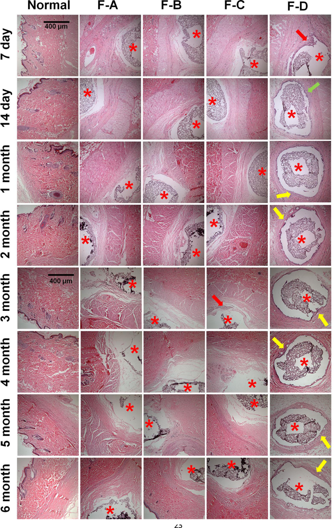
Figure 7.

In vivo pharmacodynamics of implanted composite coated dummy sensors in rats following 7-day, 14-day, 1-, 2-, 3-, 4-, 5-, and 6-months implantation (top to bottom). From the left to right columns are normal tissue, and coating-A, B, C and D, n=3 animals for each coating. Coatings-A, B, C contain dexamethasone loaded microspheres and/or free dexamethasone. Coating-D is a blank coating containing dexamethasone free microspheres. Stars indicate where the implants were located, the red arrows indicate infiltrated inflammatory cells, the green arrow indicates activated fibroblasts present during the transitional phase from acute to chronic inflammation, and the yellow arrows indicate a fibrous capsule formed around the implants.
