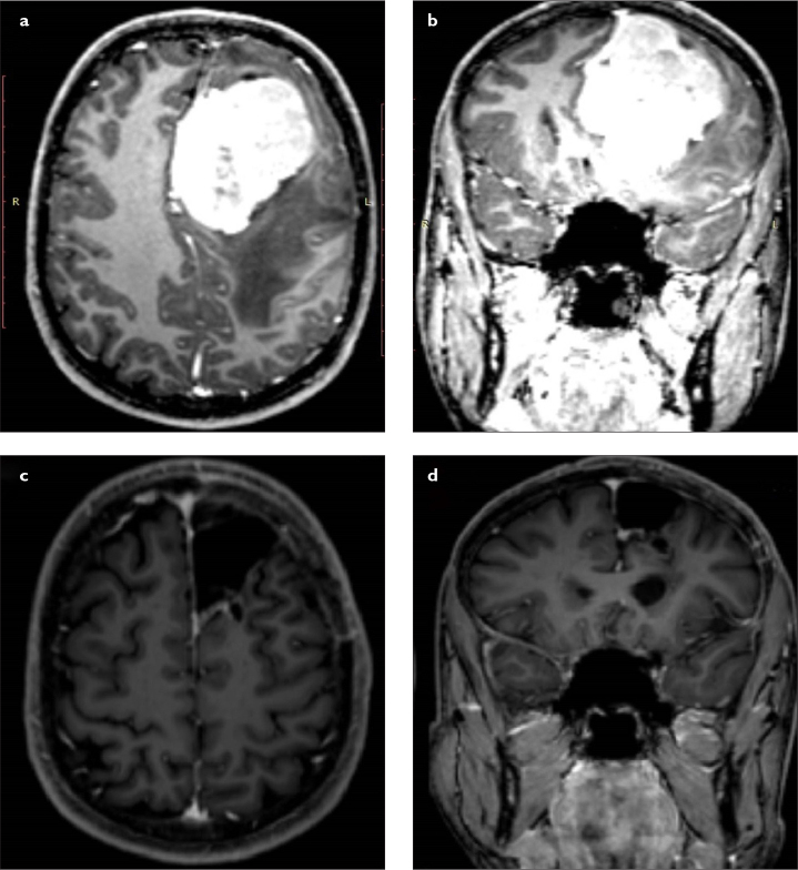
Figure 1. a–d.
(a) Preoperative axial and (b) coronal MRI scans of a patient with giant left frontal meningioma originating from the falx cerebri. The patient underwent surgical resection using left frontal craniotomy. (c) Postoperative axial and (d) coronal MRI scans confirmed gross total resection. MRI, magnetic resonance imaging.

