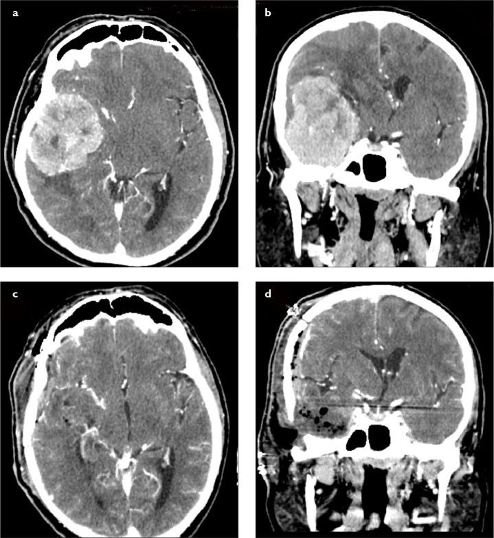
Figure 2. a–d.
(a) Preoperative axial and (b) coronal CT scans (with contrast) of a patient with giant right temporal fossa meningioma originating from the skull base dura mater. There was a significant midline shift and brain edema. The patient underwent gross total resection using right temporal craniotomy. (c) Postoperative axial and (d) coronal CT scans (with contrast) confirmed gross total resection. The shift was improved and the resolution of brain edema was obvious. CT, computed tomography.

