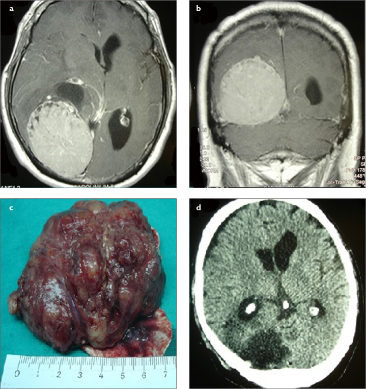
Figure 3. a–d.
(a) Preoperative axial and (b) coronal MRI scans of a patient with giant left posterior parietal meningioma. There was a significant compression on the atrium of the left lateral ventricle. The patient underwent gross total resection including dura mater using left parietal craniotomy. (c) The tumor was larger than 6 cm. (d) Postoperative axial CT scan confirmed the total resection of tumor. CT, computed tomography; MRI, magnetic resonance imaging.

