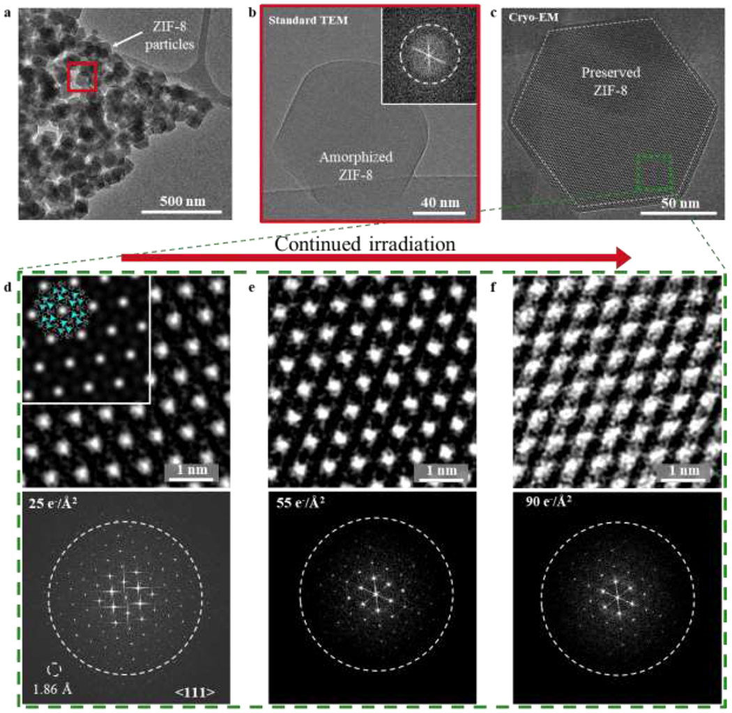
Figure 2 |. Electron irradiation of ZIF-8 at cryogenic temperatures.

a, TEM image of ZIF-8 particles taken at room temperature with electron dose rate 2 e−/Å2/s for ~1s. Diffraction contrast suggests sample is crystalline. b, HRTEM image of red boxed region from (a). After accumulated electron dose of ~50 e−/Å2, ZIF-8 becomes completely amorphous. Inset: corresponding FFT showing no crystallinity in the sample. c, Cryo-EM image of ZIF-8 (outlined by dashed white line) taken along the <111> direction at −170 °C with electron dose rate of ~4.5 e−/Å2/s for 1.5s. At 225 nm underfocus, bright spots in the ZIF-8 lattice correspond to empty pore space. d, e, f, Magnified images of green boxed region from (c) exposed to electron doses of 25 e−/Å2 (d), 50 e−/Å2 (e), and 90 e−/Å2 (f) with corresponding FFT pattern below. The dashed circle on the FFT represents an information transfer of 2.5 Å. Information transfer of 1.86 Å is possible, as indicated by the reflection circled in (d). Inset of (d): The ZIF-8 atomic structure is overlaid onto the simulated TEM image, which is calculated using 225 nm underfocus and 100 nm sample thickness. The simulated image matches reasonably well with the experimental cryo-EM image.
