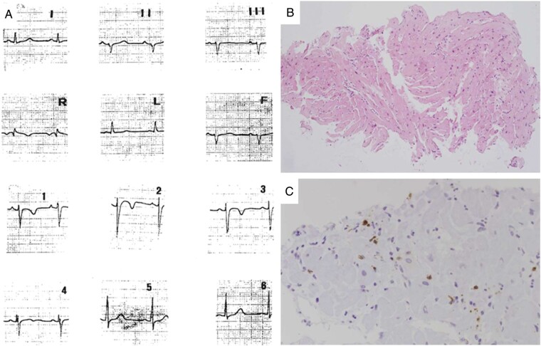
Figure 3.
A 12-year-old patient (Patient #7) with chest pain at disease onset. Twelve-lead ECG performed at the time of chest pain episode (A): note the mild ST-segment elevation in V1–V3 with T-wave inversion. Endomyocardial biopsy reveals diffuse interstitial oedema and focal inflammatory cell infiltrates (B, haematoxylin–eosin stain), which are positive for activate T-lymphocytes (C, CD3 immunohistochemistry). ECG, electrocardiogram.

