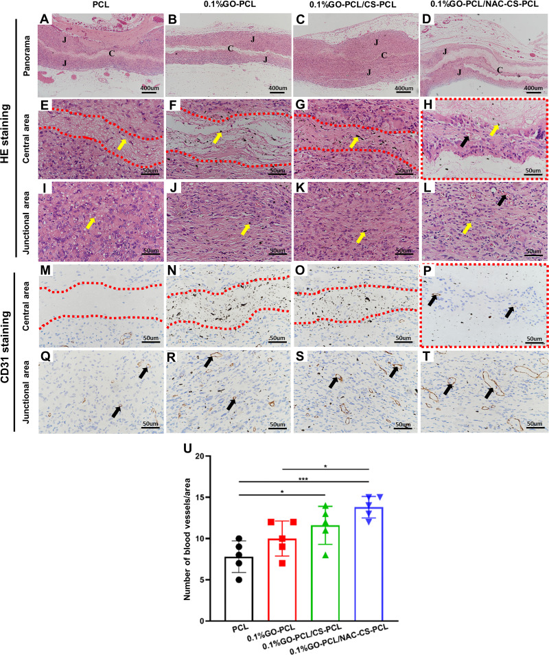
Figure 6.
Histological analysis of the scaffolds 2 months post-implantation.
Notes: (A–L) H&E staining and (M–T) CD31 immunohistochemical staining of explants at 2 months post-implantation. Capital letter C represents the central area, and capital letter J represents the junctional area. High magnification images of the central area are within the red dotted line. The yellow arrows and black arrows indicate the residual scaffold materials (E and I: PCL; F–H, J–L: GO) and new blood vessels, respectively. (U) histogram of the number of blood vessels/area (mean ± SD; *P<0.05, ***P<0.001).

