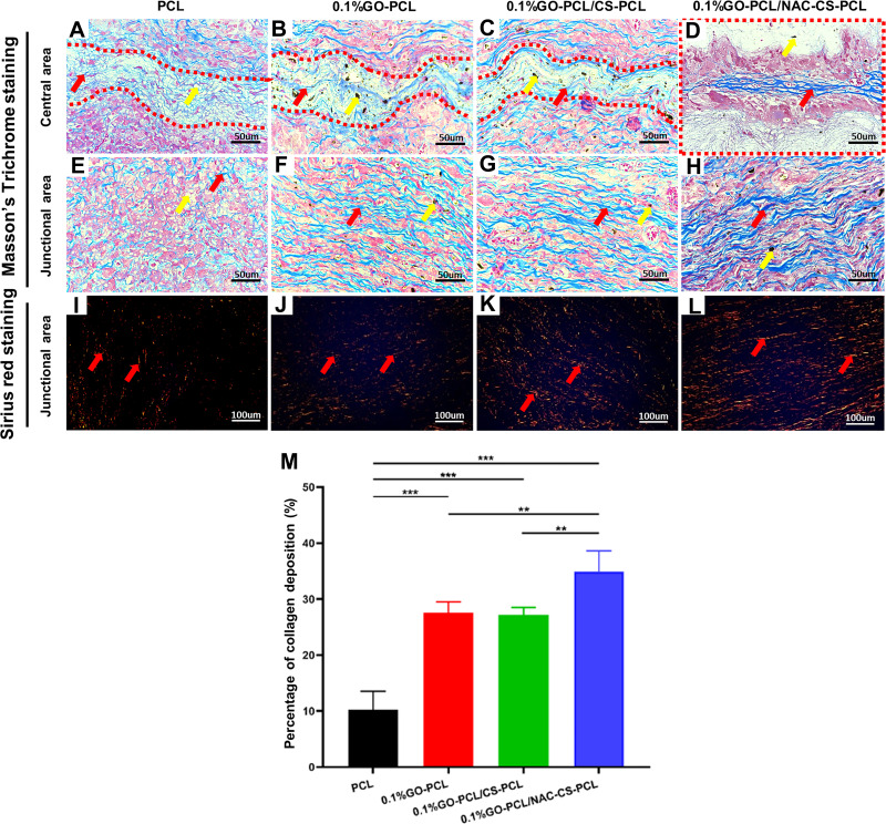
Figure 7.
New collagen deposition on the scaffolds 2 months post-implantation.
Notes: (A–H) Masson’s Trichrome staining and (I–L) Sirius red staining of explants under polarized light microscope at 2 months post-implantation. The yellow arrows and red arrows indicate the residual scaffold materials (A and E: PCL; B–D, F–H: GO) and new collagen tissues, respectively. (M) histogram of the percentage of collagen deposition per area (mean ± SD; **P<0.01, ***P<0.001).

