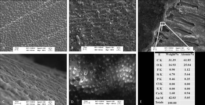
Fig. 3.
Scanning electron micrographs of the morphological analysis of the surface and cross-sectional areas of the dentin: ( A ) shows a representative scanning electron micrograph of the demineralized dentin with open tubules. ( B ) shows a representative scanning electron micrograph of the dentin immediately after brushing with the toothpaste with occluded dentinal tubules. ( C ) shows the dentin surface in which a mineralized layer was formed after 7 days of treatment. ( D ) shows the scanning electron micrograph of the treated dentin with a majority of occluded tubules after 1 week of treatment. ( E ) shows a scanning electron micrograph of the mineralized layer formed at the dentin surface, demonstrating the presence of silicon (6.70 weight%), fluorine (0.90 weight%), calcium (1.60 weight%), and phosphorus (0.46 weight%) in the elemental analysis.

