
FIG. 4.
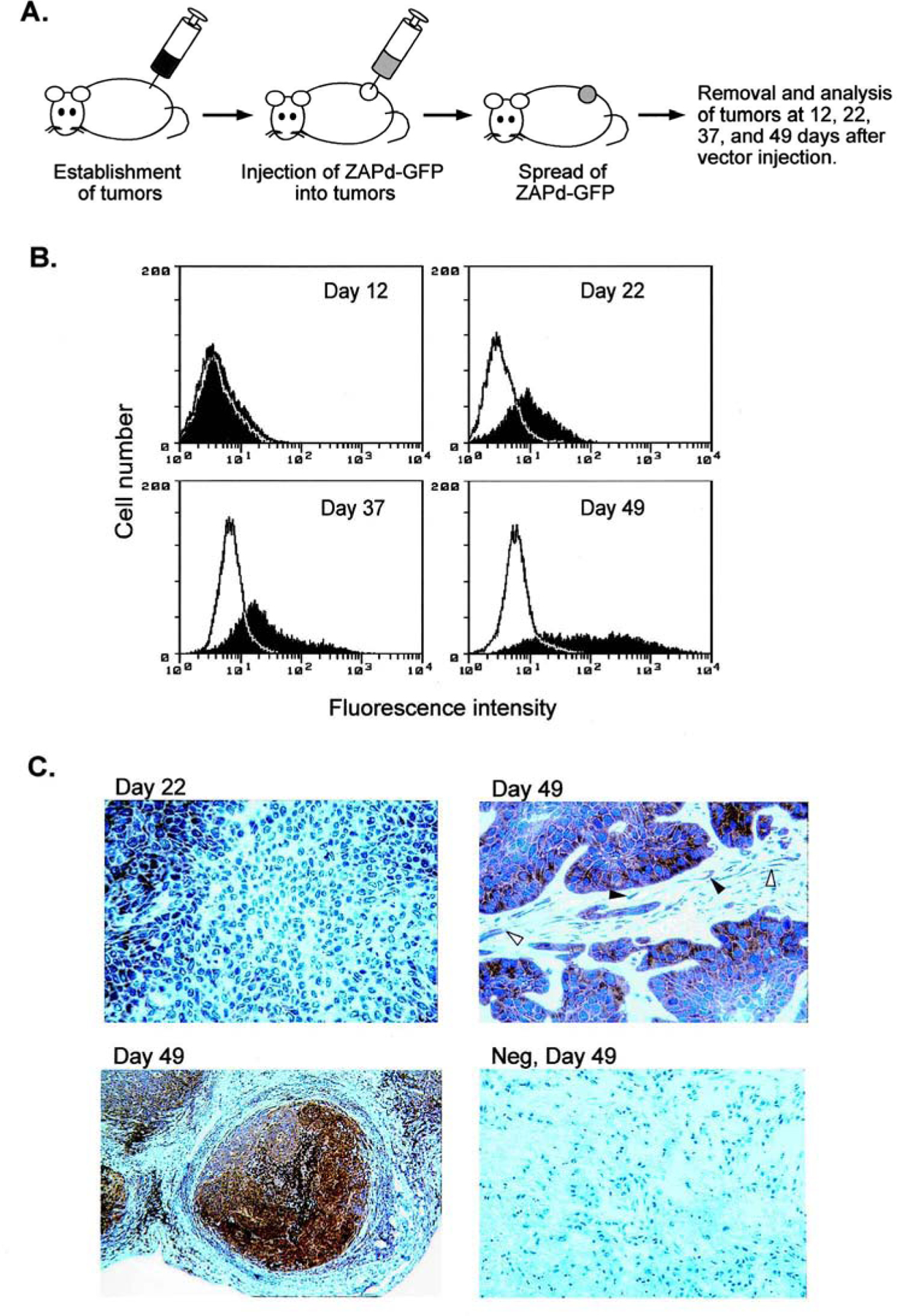
Spread of ZAPd-GFP through solid tumors in mice. (A) Subcutaneous tumors, 1 to 1.5 cm3 in volume, were injected with 6 × 10³ PFU of ZAPd-GFP. Twelve, 22, 37, and 49 days after vector injection, tumors were removed from subsets of the mice and were analyzed for virus spread by flow cytometry and Southern blot hybridization. (B) Expression of GFP transgene in tumors. Tumor cells from each of the four time points were dissociated into single-cell suspensions and analyzed for GFP fluorescence by flow cytometry. Shaded histograms represent tumors injected with ZAPd-GFP, and open histograms represent untreated tumors. (C) Immunohistochemical staining of GFP in tumors injected with ZAPd-GFP. Tumors removed 22 and 49 days after vector injection were stained with a monoclonal antibody to GFP and counterstained with hematoxylin. Top left: Tumor removed 22 days after vector injection. Top right and bottom left: Tumors removed at 49 days. Open arrowheads indicate transduced fibroblasts and closed arrowheads indicate transduced endothelial cells. Bottom right: Negative control tumor removed 49 days after vector injection. Each panel represents a different mouse. Original magnification: Top left and bottom right, ×300; top right, ×400; bottom left, ×6.