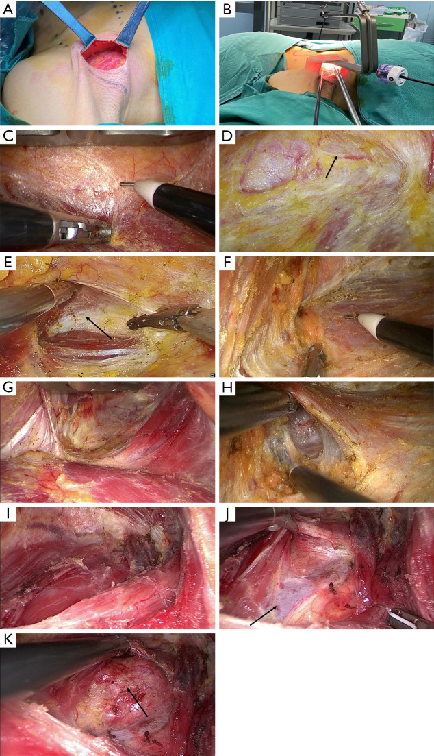
Figure 2.
Creation of a surgical cavity. (A) Exposure of the lateral edge of the pectoralis major muscle. (B) Placement of operating instruments. (C) Separation of the pectoralis major myocutaneous flap. (D) Protection of the supraclavicular cutaneous nerve (arrow shows the cutaneous nerve). (E) Identification of the sternocleidomastoid muscle gap (arrow shows the muscle gap). (F) Identification of the posterior edge of sternocleidomastoid muscle. (G) Separation of the sternocleidomastoid muscle gap. (H) Separation of the posterior edge of sternocleidomastoid muscle. (I) Dissection of the omohyoid muscle (arrow shows the omohyoid muscle). (J) Exposure of the “muscular triangle” at the junction between the lateral side of strap muscle and the omohyoid muscle (arrow shows the internal jugular vein). (K) Traction of the strap muscle to expose the thyroid (the arrow shows the thyroid).

