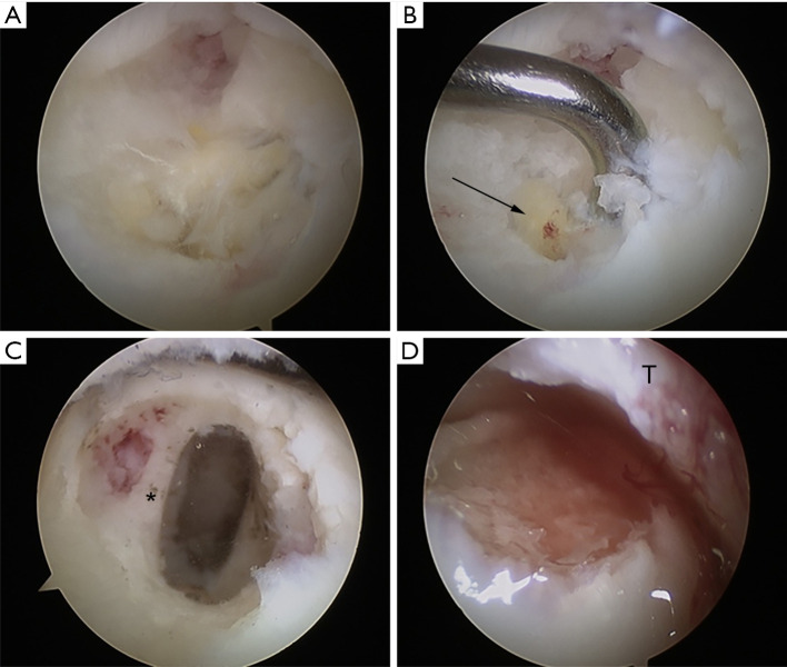
Figure 1.
Intraoperative images. (A) OLT was confirmed and exposed. (B) Detection of the OLT with the probe. The endoscopic view shows an OLT with a subchondral cyst (black arrow). (C) Endoscopic view after debridement and scraping of the OLT shows the healthy bone bed (*). (D) Lesion site after bone graft transplantation and fixation with fibrin glue. T, tibia; OLT, osteochondral lesion of the talus.

