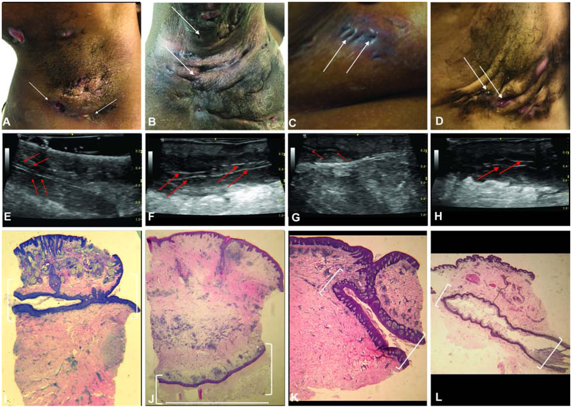
Figure 1: Ultrasonography identifies deep dermal tunnels in HS.

Clinical assessment of tunnels marked by superficial ostia (white arrows) (A) Axilla (B) Axilla (C) Breast (D) Axilla. (E-H) Corresponding ultrasound images of tunnels detected by clinical examination. Red arrows highlight the hyperechoic border of the tunnel on ultrasound. (E-H) Light microscopy of the tunnel (1.2x magnification). White brackets outline the tunnel.
