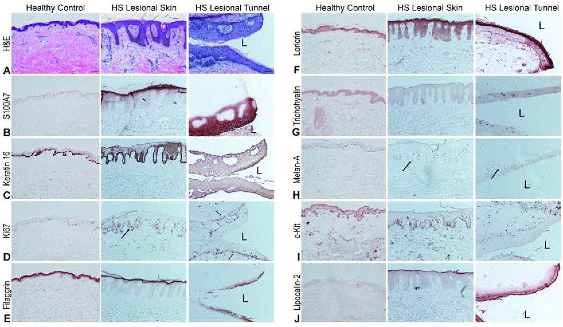
Figure 2: HS tunnels recapitulate the structural properties of the overlying epidermis.

Representative biopsies from HS patients and site-matched healthy volunteers stained with (A) H&E demonstrating prominent psoriasiform lengthening of the rete ridges, thinning of the suprapapillary plate, hyper and parakeratosies as well as reduction of the granular layer in the HS epidermis compared to healthy controls. HS tunnels contain a thick stratified squamous epithelium with increasing differentiation towards the lumen (L). Scale Bar, 100μm (B) S100A7 positivity (C) Keratin-16 and (D) Ki67 identify this epithelium as composed of dividing keratinocytes with increasing differentiation towards the luminal layer compared to the more basal cells (D, black arrows). Differentiation is indicated by filaggrin (E) and loricrin (F) staining. Intermittent positive trichohyalin staining (G) is also observed. Other cell types within the tunnel include melanocytes (H) with c-Kit identifying dermal mast cells (I). (J) Lipocalin-2 staining is also increased in intensity in the luminal layers of the tunnel epithelium compared to superficial HS epithelium.
