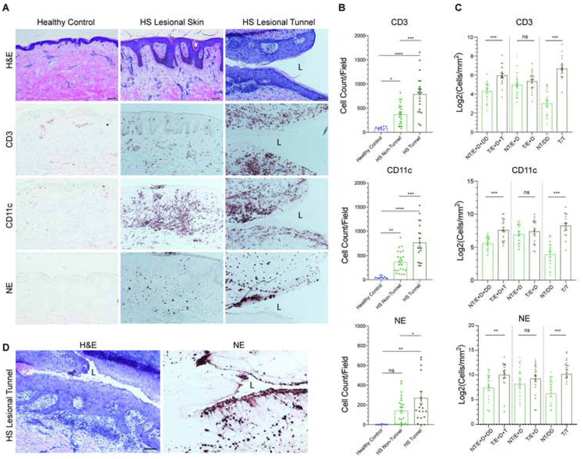
Figure 3: Tunnels are immunologically active.

(A) Immunohistochemistry demonstrates increased infiltration of CD3, CD11c, and NE positive cells in HS compared to site-matched healthy controls. Epidermotropism and transepithelial migration towards the tunnels are also observed. Scale Bar, 100μm. “L” denotes lumen. (B) Quantitative CD3+, CD11c+ and NE+ cell counts highlights a significant difference in CD3, CD11c and NE positive cells between HS samples with and without tunnels. (C) Density of CD3, CD11c, and NE positive cellular infiltrate was analyzed within non-tunnel (NT) and tunnel (T) HS specimens stratified by location of cells within the biopsy (E=Epidermis; D=Dermis; T=Tunnel and depth-matched DD=Deep Dermis). There is a significant increase in inflammatory infiltration between tunnel and non-tunnel specimens when the deep dermal component of biopsies is taken into account. No significant elevation of CD3+ CD11c+ and NE+ cell density was seen between the epidermis and the superficial dermis in tunnel and non-tunnel specimens. Results are the mean ± SEM *p<0.05, **p<0.01, ***p<0.001 (D) Dense clusters of neutrophils undergoing NETosis in the tunnel epithelium adjacent to the lumen (L).
