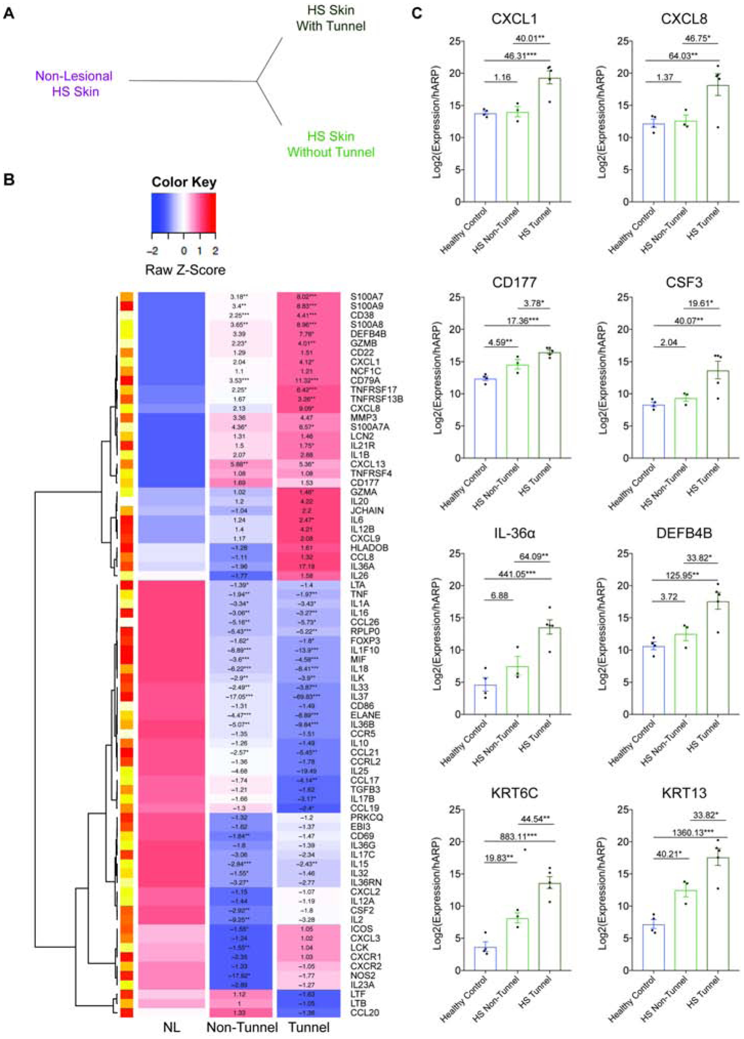
Figure 4: HS samples cluster based on presence of tunnels.

(A) Unsupervised hierarchical clustering analysis of TLDA data based on the histological presence of tunnels demonstrates distinct clustering of tunnel and non-tunnel biopsy specimens compared to non lesional tissue (B) Heatmap of differential gene expression of HS-associated genes in HS Non-Lesional (NL) specimens (n=7), HS samples without tunnels (n=10) and HS samples containing tunnels (n=6), all confirmed by histological presence of tunnel. Results indicate FCH *p<0.05, **p<0.01, ***p<0.001. (C) Confirmatory RT-PCR of healthy controls (n=4), and actively inflamed HS lesional samples without (n=3), and with tunnels (n=5). Results are the mean ± SEM, FCH is shown. *p<0.05, **p<0.01, ***p<0.001
