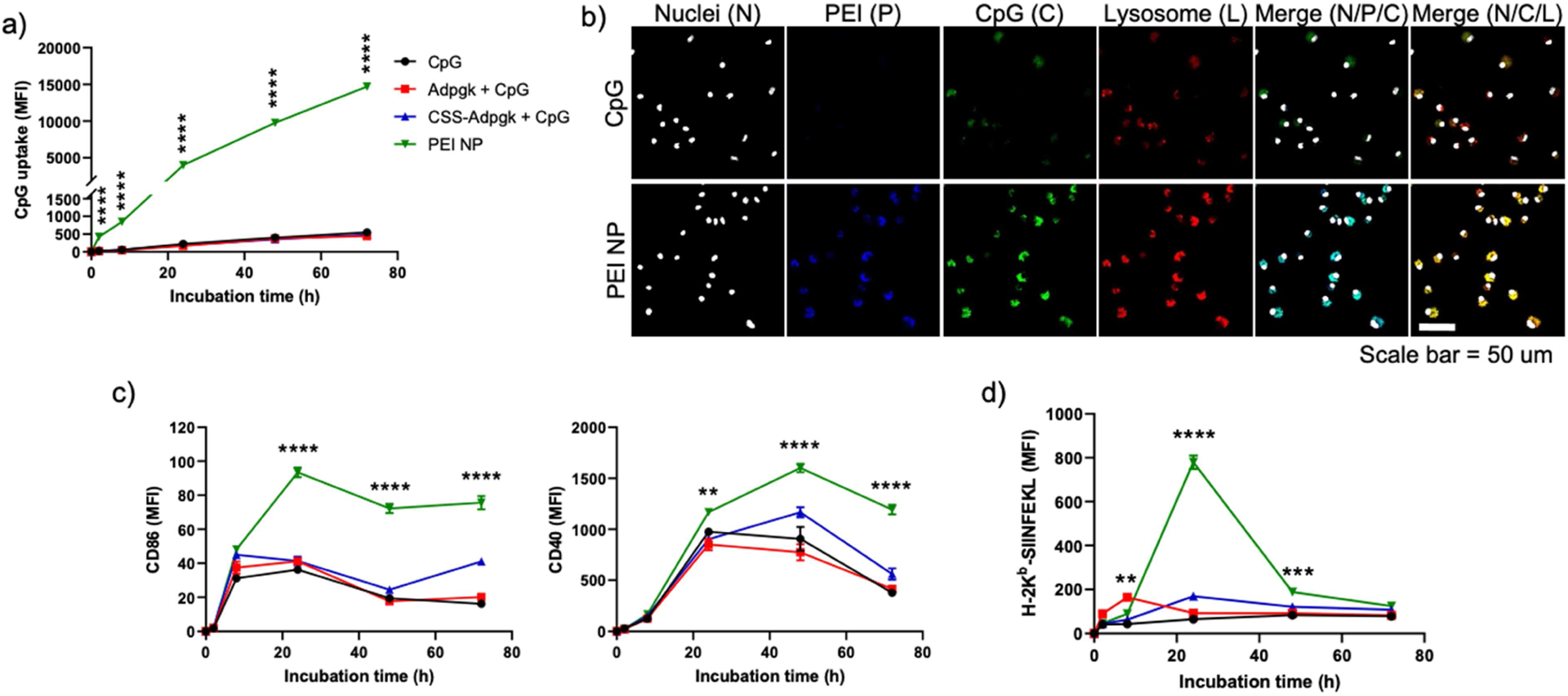
Figure 2. NP Vacc promotes cellular uptake of antigen and CpG by DCs and improves DC maturation and antigen cross-presentation.

a-b) BMDCs were incubated in vitro with fluorophore-labeled CpG in the formulations of CpG, Adpgk + CpG, CSS-Adpgk + CpG, or corresponding NP VACC, and fluorescence signals were measured using a) flow cytometry and b) confocal microscopy (scale bar = 50 μm). c-d) BMDCs were incubated with CpG, SIINFEKL + CpG, CSS-SIINFEKL + CpG, or NP Vacc, and c) DC activation and d) antigen presentation were measured by staining cells with c) anti-CD86 and anti-CD40 antibodies or d) anti-H-2Kb-SIINFEKL antibody, respectively, followed by flow cytometry. Data are presented as mean ± SEM. *p < 0.05, **p < 0.01, ***p < 0.001, and ****p < 0.0001, analyzed by two-way ANOVA, followed by Tukey’s HSD multiple comparison post hoc test. Asterisks represent comparison between NP Vacc vs. Adpgk + CpG in a), and between NP Vacc vs. SIINFEKL + CpG in c) and d).
