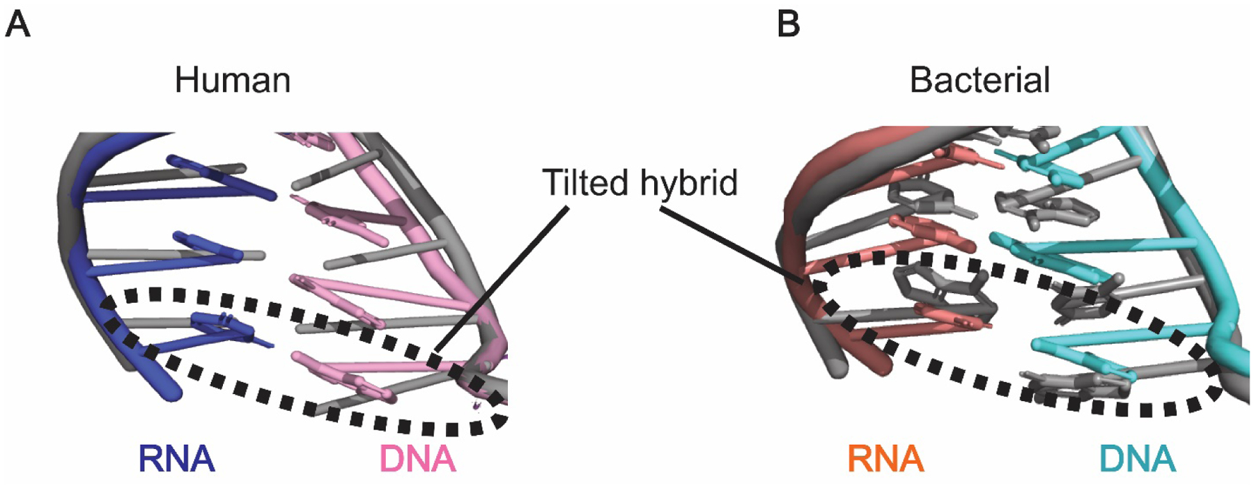
Figure 3: Half-translocated RNA-DNA duplex in the paused RNA polymerase active site.

A) The RNA-DNA duplex observed in the Pol II-DSIF-NELF structure (colored, PDB ID: 6GML) is in a “tilted” conformation relative to the RNA-DNA duplex observed in the Pol II-DSIF structure (grey, PDB ID: 5OIK). B) The RNA-DNA hybrid in the active site of the paused bacterial RNA polymerase (colored, PDB ID: 6ASX) adopts a similarly tilted conformation compared to the hybrid in the active site of a post-translocated RNA polymerase (grey, PDB ID: 6ALF).
