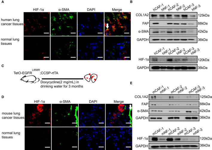
FIGURE 1.

HIF‐1α is highly expressed in CAFs of LC. A, Representative immunofluorescence images of HIF‐1α (red) and α‐SMA (green) in the frozen sections from human lung cancer tissues and normal lung tissues (Scale bar, 50 μm). B, The protein levels of COL1A2, FAP, α‐SMA and HIF‐1α were determined by western blot from hCAFs and their counterpart hNFs isolated from three lung cancer patients. C, The doxycycline‐induced mouse spontaneous lung cancer model (TetO‐EGFRL858R; CCSP‐rtTA) was established. D, Representative immunofluorescence images of HIF‐1α (red) and α‐SMA (green) in the frozen sections from mouse lung cancer tissues and normal lung tissues (Scale bar, 50 μm). E, The protein levels of COL1A2, FAP, α‐SMA and HIF‐1α were determined by western blot from mCAFs and mNFs isolated from mouse lung cancer tissues and lung normal tissues
