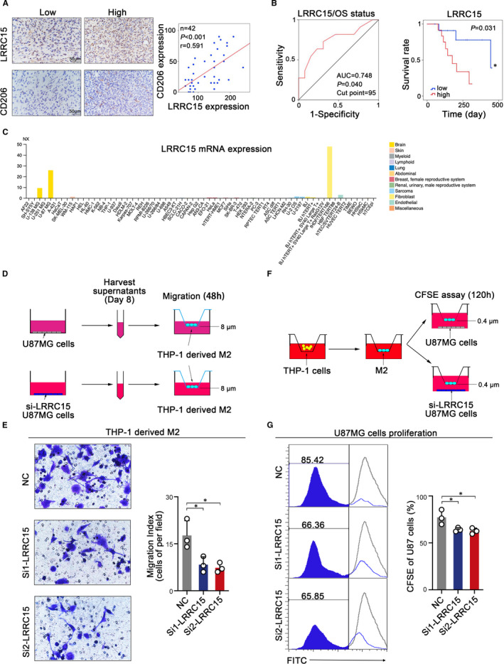
FIGURE 5.

LRRC15 promotes the recruitment of GAMs and proliferation of tumour cells. (A) Representative micrographs of LRRC15 expression and CD206 expression as well as their expression correlation analysis in human rGBM tissues by immunohistochemistry assay. (B) Kaplan‐Meier curve showed the association between LRRC15 expression and OS in 24 rGBM patients with survival information based on the relative LRRC15 IHC score divided by the optimal cut‐point off. ROC curve analysis of LRRC15 IHC score was shown based on OS status. (C) LRRC15 expression in multiple cells from Human Protein Atlas. (D) Schematic diagram of experimental design of tumour LRRC15 expression influencing M2‐like macrophage migration. Supernatants of tumour cell culture were collected on Day 8 after the tumours were seeded and used for the M2‐like macrophages migration assay. (E) Migration analysis of M2‐like macrophages in the presence of LRRC15 or LRRC15 knockdown tumour supernatants by transwell assay. Images were taken 48 h after the M2‐like macrophages were exposed to the tumour supernatants. The images represent 1 of 3 individual experiments using THP‐1‐derived macrophages. (F) Schematic diagram of experimental design of tumour LRRC15 expression influencing tumour proliferation in the presence of M2‐like macrophages. THP‐1 cells were induced into M2‐like macrophages and co‐cultured with U‐87 MG cells with/without LRRC15 knockdown for CFSE assay (G) CFSE assay was used to examine the tumour cells proliferation with/without LRRC15 knockdown
