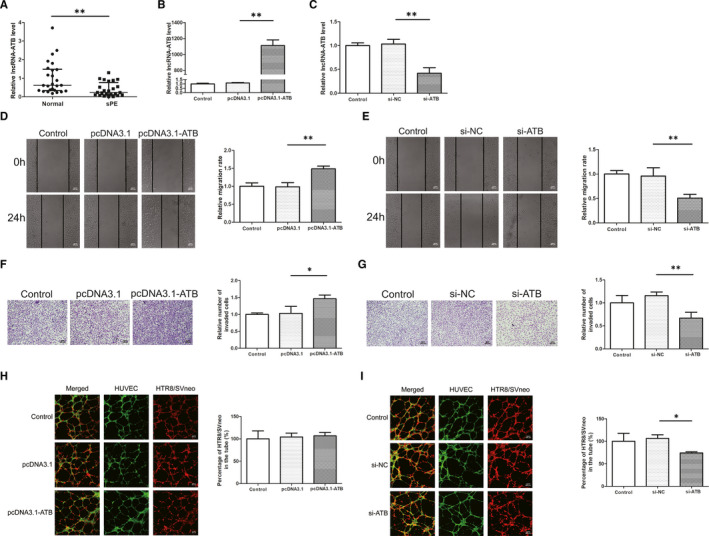
FIGURE 2.

Expression of lncRNA‐ATB in PE tissues and effects on cell migration, invasion, and co‐culture model. A, The quantitative real‐time PCR results showed that the expression of placental lncRNA‐ATB in PE patients was lower than that in normal pregnant women. B, C, The expression of lncRNA‐ATB was significantly increased in HTR8/SVneo transfected with pcDNA3.1‐ATB and decreased with si‐ATB compared to control groups. D, E, Overexpression of lncRNA‐ATB in HTR8/SVneo cells resulted in increasing wound closure ability and lncRNA‐ATB knockdown reduced the rate of wound closure. Original magnification×100. The scale bars indicate 500 μm. F, G, Overexpression of lncRNA‐ATB significantly increased cell invasion while knockdown of lncRNA‐ATB reduced cell invasion compared with the control group. Original magnification×100. The scale bars indicate 200 μm. H, I, HTR8/SVneo cell integration into endothelial cellular networks was significantly inhibited by si‐ATB treatment while lncRNA‐ATB overexpression did not affect the integration. Original magnification×100. The scale bars indicate 500 μm. *P < .05, **P < .01
