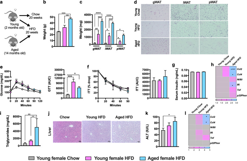
Fig. 2. HFD amplifies aging-associated weight gain and uncovers aging-dependent skewing of metabolic disease severity in female mice.
2-month-old (Young) and 14-month-old (Aged) female C57BL/6 mice, (n = 3–5/condition) were housed at 22 °C (TS) and fed Chow or HFD for up to 20 weeks. a Schematic representation of DIO model used. b Body weight at time of harvest. c White adipose tissue (WAT) weight: gonadal WAT (gWAT), inguinal WAT (iWAT) and perirenal WAT (pWAT). d gWAT, iWAT and pWAT hematoxylin eosin (H&E) staining. e Left, glucose tolerance test (GTT) and Right, GTT area under the curve (AUC). f Left, insulin tolerance test (ITT) and Right, ITT area under the curve (AUC). g Serum insulin levels. h Liver triglycerides. i Liver H&E staining. j Serum Alanine transaminase (ALT) levels. k Heatmap of WAT mRNA levels of Ccl2, Ccl4, f4/80, Il6, Tnf and p22phox fold change relative to chow diet fed young female. l Heatmap of hepatic mRNA levels of Ccl2, Ccl4, f4/80, Il6, Tnf and p22phox. A representative of 2 individual experiments. Data represent means + SE. b, c, e–i, k–m. One-way ANOVA *P < 0.05, **P < 0.005 and ***P < 0.0005.

