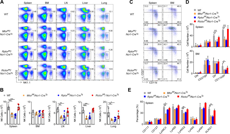
Fig. 6. Terminal differentiation of NK cells requires mTORC1 rather than mTORC2.
Representative flow cytometric profiles (A) and the absolute number (B) of NK cells in the spleen, bone marrow (BM), lymph nodes (LN), livers, and lungs from WT (n = 6), Mtorfl/fl/Ncr1-CreTg (n = 4), Rptorfl/fl/Ncr1-CreTg (n = 5), and Rictorfl/fl/Ncr1-CreTg (n = 5) mice are illustrated. Representative flow cytometric profiles (C) and enumeration (D) of NK cell subsets in the spleen and BM from the WT (n = 6), Mtorfl/fl/Ncr1-CreTg (n = 4), Rptorfl/fl/Ncr1-CreTg (n = 5), and Rictorfl/fl/Ncr1-CreTg (n = 5) mice are illustrated. E Flow cytometry analysis of development-related NK cell receptors of CD3−NK1.1+ cells in the spleen is illustrated (n = 2–3/genotype). For the bar graphs, each dot represents one mouse. All data represent three independent experiments, and one (E) or two-pooled (B, D) independent experiments are illustrated. All data are indicated as mean ± SD. Statistical significance was calculated using one-way ANOVA. *P < 0.05, **P < 0.01, and ***P < 0.001.

