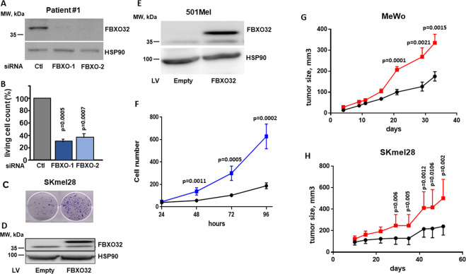
Fig. 4. FBXO32 controls proliferation and xenograft development.
A Western blot analysis of FBXO32 expression after transfection of siRNAs targeting FBXO32 in melanoma cells isolated from patient 1. HSP90 expression was probed as loading control. B Quantification of living cells 72 h after FBXO32 downregulation by siRNAs in melanoma cells from patient 1 (mean ratio ± SD, n = 3). C Colony formation assay of parental or FBXO32 overexpressing SKmel28 cells. D Western blot analysis of FBXO32 expression after FBXO32-forced expression in SKmel28 cells. HSP90 expression was probed as loading control. E Western blot analysis of FBXO32 expression after lentivirus-mediated FBXO32-forced expression in 501Mel cells. HSP90 expression was probed as loading control. F Quantification of 501Mel cell proliferation after empty virus (black line) or FBXO32 encoding virus (blue line) transduction, from 24 to 96 h (mean ± SD, n = 6). G Xenograft growth after subcutaneous injection of MeWo cells transduced with empty vector (black line) or FBXO32 encoding virus (red line) (mean ± SD, n = 10). H Xenograft growth after subcutaneous injection of SKmel28 cells transduced with empty vector (black line) or FBXO32 encoding virus (red line) (mean ± SD, n = 10).

